FLUX AI Image générée par Flux1.1 Pro
Rapide
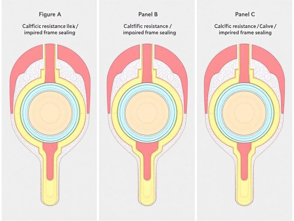
Create a professional, publication-ready, three-panel scientific illustration for a cardiology review article in Journal of Clinical Medicine (MDPI style). Figure title: "Figure 1. Three mechanistic pathways of paravalvular leak after transcatheter aortic valve implantation (TAVI)" Style: Clean, schematic medical textbook illustration, vector-style, high scientific accuracy, not photorealistic. Clean black outlines, subtle shading, flat medical colors with depth. White background. MDPI / Nature Medicine level quality. Publication-ready, high resolution. Layout: Three equal vertical panels (A, B, C) arranged side-by-side horizontally, clearly labeled at the top of each panel in bold: "Panel A", "Panel B", "Panel C". Professional spacing, balanced composition. --- **Panel A – Mechanism 1: Calcific resistance / impaired frame sealing** Cross-sectional (short-axis) view at the level of the aortic annulus. Left side: perfectly circular THV frame (light blue) with excellent apposition and sealing skirt (gray) against pink aortic annulus tissue. Right side: same frame deformed by a large bulky calcific protrusion (8–12 mm, white/cream with hard shadow) originating from the non-coronary or left coronary cusp. Visible gap between the skirt and tissue. Prominent red arrow labeled "calcific protrusion". Small inset sagittal view on the right showing how the sealing skirt cannot fill the space behind the calcium nodule. Label below panel: "Calcific resistance preventing complete frame apposition" --- **Panel B – Mechanism 2: Geometric mismatch (elliptical annulus)** Top-down (short-axis) view of a clearly elliptical aortic annulus (EI ≈ 0.65, approximate dimensions 25 × 19 mm). Circular THV frame expanded inside the ellipse. Bright red zones at the minor axis poles indicating paravalvular leak gaps. Label: "Effective undersizing at minor axis poles despite nominal area-based sizing" Small comparative inset on the right: self-expanding valve with conformable outer skirt adapting better to the elliptical shape versus balloon-expandable valve. Label below panel: "Geometric mismatch due to elliptical annulus" --- **Panel C – Mechanism 3: LVOT calcium / suboptimal seating depth** Sagittal long-axis view through the LVOT, aortic annulus and ascending aorta. Prominent subannular calcification (grade 2–3, white/cream) on the septal side acting as a physical barrier. Two THV positions shown: 1. Implanted too high – skirt does not cover the lower annular border (arrow: "LVOT calcium acting as physical stop") 2. Implanted too deep – excessive protrusion into LVOT Grey shaded zone indicating narrowed "optimal implantation depth window" constrained by LVOT calcium. Label below panel: "Suboptimal seating depth caused by LVOT calcification" --- Technical requirements: - Extremely clean lines, precise anatomical proportions, readable sans-serif labels (Arial-like) - Medical color palette: light blue (THV frame), gray (skirt), pink-red gradient (myocardium/aorta), white/cream with shadow (calcification), bright red (leak areas) - Clear arrows with labels - High scientific accuracy and visual clarity - MDPI-compatible style, suitable for print at 300 dpi - Add main figure caption at the bottom: "Three mechanistic pathways of paravalvular leak after TAVI"


